Fig. 1.
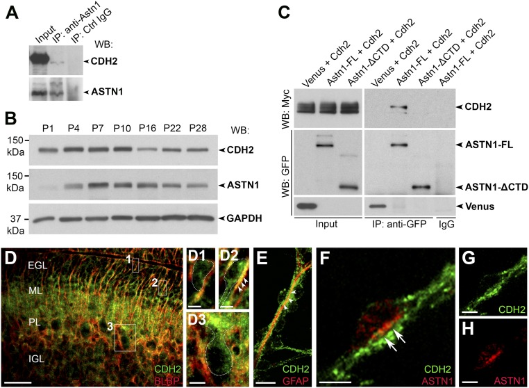
ASTN1 and CDH2 form cis interactions and colocalize in the migration junction. (A) In vivo immunoprecipitation of ASTN1 in P7 whole-cerebellar lysates blotted with ASTN1 and CDH2 antibodies. ASTN1 is part of a protein complex with CDH2. (B) Developmental protein expression of CDH2 and ASTN1 in the cerebellum of postnatal mice (P1–P28) by Western blot. CDH2 expression was highest between P4–P10, decreased by P16, and reached a steady level at P22–P28. ASTN1 expression increased after P1 and was highest at P7–P10. Protein expression was compared with GAPDH levels. (C) Western blots showing coimmunoprecipitation of ASTN1-Venus and CDH2-Myc in HEK 293T cells. CDH2 interacted with ASTN1-FL but not with ASTN1-ΔCTD. (D and E) Endogenous protein expression of CDH2 at P7 in sagittal mouse cerebellar sections (D) and in GCP/BG in vitro cocultures (E). CDH2 was expressed in GCPs in the EGL (D1), in migrating GCPs in the ML (D2), and in Purkinje cells (D3) and colocalized with BLBP and GFAP in BG fibers (arrowheads in D2 and E). (F–H) GCP/BG cocultures labeled with antibodies against CDH2 (F and G) and ASTN1 (F and H). CDH2 localized to neuronal processes, glial fibers, and the migration junction beneath the neuronal soma. ASTN1 colocalized with CDH2 in the migration junction (arrows in F). PL, Purkinje cell layer. (Scale bars: 50 µm in D; 5 µm in D1, D2, and F–H; and 10 µm in D3 and E.)

