Fig. 3.
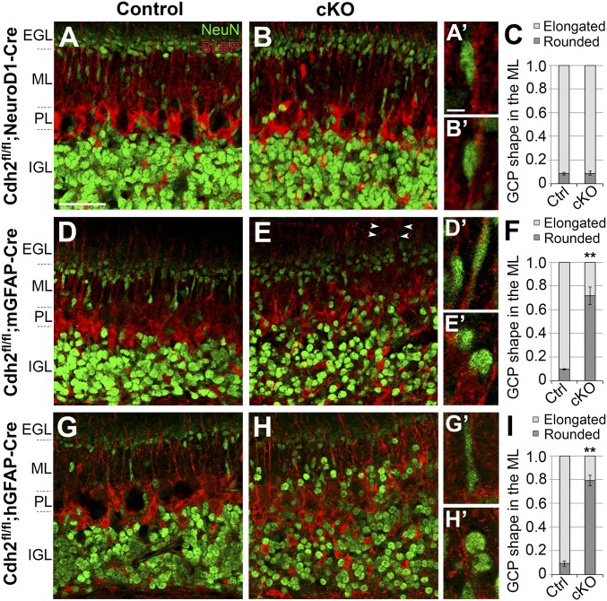
Neuronal migration in Cdh2-cKO mice. Sagittal cerebellar sections of P7 Cdh2fl/fl control mice (A, D, and G) and Cdh2-cKO littermates expressing NeuroD1-Cre (B), mGFAP-Cre (E), or hGFAP-Cre (H) and labeled with NeuN and BLBP antibodies. In control mice, NeuN-positive GCPs displayed elongated somas along BG fibers in the ML, indicating migrating cells. A similar phenotype is seen in mice with GCPs lacking Cdh2 (B). In contrast, a loss of Cdh2 in BG (E) or in both GCPs and BG (H) resulted in rounded GCPs and a stalled migration in the ML. The proportion of elongated and rounded GCPs in the ML is shown in stacked bar charts (C, F, and I). Abnormal radial patterning of BG fibers was observed in some areas (arrowheads in E). Higher magnifications in A′, B′, D′, E′, G′, and H′ show representative GCPs from each genotype. PL, Purkinje cell layer. **P < 0.01. (Scale bars: 50 µm in A, B, D, E, G, and H and 5 µm in A′, B′, D′, E′, G′, and H′.)

