Figure 4.
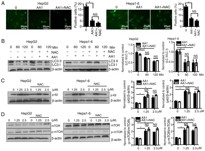
Targeting TrxR inhibits autophagy of liver cancer cells in a reactive oxygen species-independent manner. HepG2 or Hepa1-6 cells were infected with Ad-mCherry-green fluorescent protein-LC3B for 48 h and following pretreatment with NAC for 2 h or no treatment, cells were then treated with 10 µM AA1 for 6 h. (Aa) Autophagosomes were imaged by fluorescence microscopy (scale bar 20 µm), quantitative analysis of LC3 expression was presented in (Ab). (B) LC3-II and LC3-I levels were examined by western blotting following treatment with AA1 (10 µM), AA1+NAC pretreated, or NAC alone for different time points in Hepa1-6 and HepG2 cells. (C) The levels of p62 protein were detected following treatment with an increasing concentration (0–2.5 µM) of AA1 for 3 h in Hepa1-6 and HepG2 cells with or without NAC pretreatment. (D) Activation of mTOR in HepG2 and Hepa1-6 cells treated as described above was detected by western blotting. The western blots are presented on the left and quantitative analysis is presented in the right panel. Experiments were run in triplicate. *P<0.05 vs. the untreated group (0 µM). mTOR, mammalian target of rapamycin; LC3, light chain 3; NAC, N-acetylcysteine; AA1, chloro(triethylphosphine)gold(I); TrxR, thioredoxin reductase; NS, not significant.

