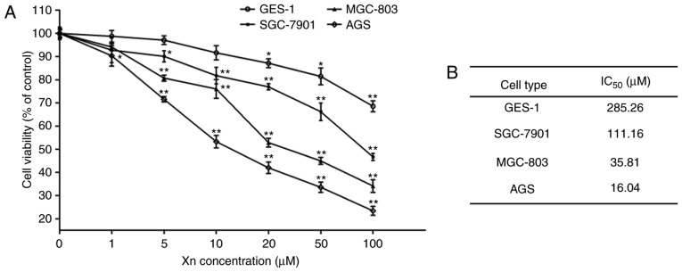
Figure 2.
Cytotoxicity of xanthohumol (Xn) on GC cells and normal gastric epithelial cells. (A) GC cells (MGC-803, SGC-7901 and AGS) and normal gastric epithelial cells (GES-1) were treated with Xn at concentrations of 0–100 µM for 24 h. (B) The IC50 for each cancer cell line was calculated according to the data in (A). Cell viability was determined by the MTS assay. Treatment with 0 µM Xn was used as control. Data are expressed as mean ± standard error of the mean. n=3. *P<0.05, **P<0.01 vs. control (0 µM Xn).

