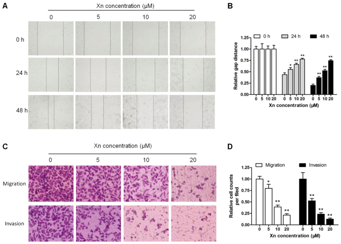
Figure 5.
Effect of xanthohumol (Xn) on wound healing, migration and invasion of AGS cells. (A and B) Cells were treated with different concentrations of Xn (0–20 µM) for 0, 24 and 48 h, followed by measurement of the relative wound width. (C and D) The Transwell assay was performed to evaluate cell migration and invasion ability; cells were treated with Xn as mentioned above in culture wells for 24 h, and the migrating and invading cells were stained by crystal violet solution. Data are expressed as mean ± standard error of the mean. n=3. *P<0.05, **P<0.01 vs. control (0 µM Xn).

