Figure 5.
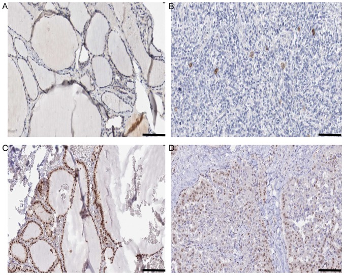
Histology information of KIT and SMAD9 in N vs. C comparison. (A) Expression of KIT in N Antibody (cat. no., HPA004471; Sigma-Aldrich; Merck KGaA, Darmstadt, Germany). Sex, female; age, 22 years; Patient ID, 2146. Glandular cells Staining: Low Intensity. Weak Quantity: 75–25%. Location: Cytoplasmic/membranous. Website: http://www.proteinatlas.org/ENSG00000157404-KIT/tissue/thyroid+gland#img. (B) Expression of KIT in C Antibody (cat. no., HPA004471; Sigma-Aldrich; Merck KGaA). Sex, female; age, 77 years; Patient ID, 2479. Staining: Not detected. Intensity: Negative. Quantity: Negative. Location: None. Website: http://www.proteinatlas.org/ENSG00000157404-KIT/cancer/tissue/thyroid+cancer#img. (C) Expression of SMAD9 in N Antibody. (cat no., HPA031162; Sigma-Aldrich; Merck KGaA). Sex, female; age, 39 years; Patient ID, 1948. Staining: High. Intensity: Strong. Quantity: >75%. Location: Nuclear. Website: http://www.proteinatlas.org/ENSG00000120693-SMAD9/tissue/thyroid+gland#img. (D) Expression of SMAD9 in C Antibody. (cat. no., HPA031162; Sigma-Aldrich; Merck KGaA). Sex, male; age, 75 years; Patient ID, 3107. Staining: Medium. Intensity: Moderate. Quantity: >75%. Location: Nuclear. Website: http://www.proteinatlas.org/ENSG00000120693-SMAD9/cancer/tissue/thyroid+cancer#img. Scale bar, 100 µm.

