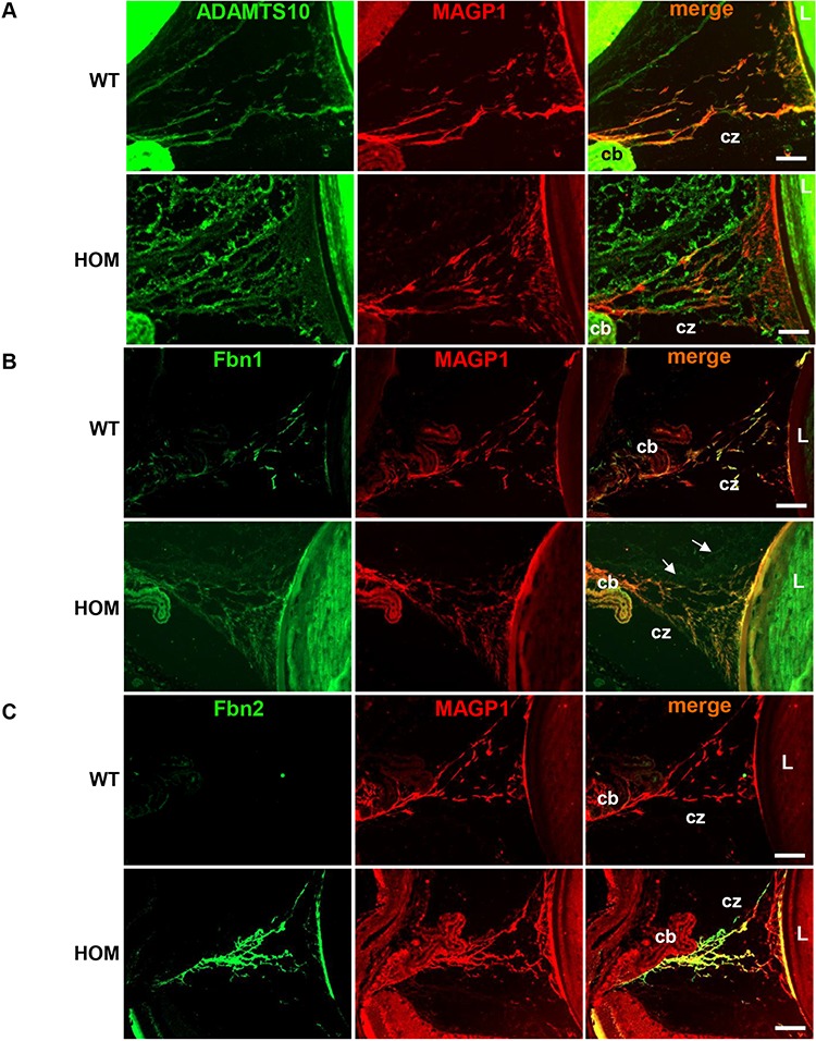
Figure 5.

Secretion of mutant ADAMTS10 and increased expression of Fbn2 in the CZs. Immunohistochemical staining in the 3-month-old WT and HOM CZ of (A) ADAMTS10 (green) and MAGP1 (red) showing reduced co-localization in the mutant samples. (B) FBN1 (green) and MAGP1 (red) fully co-localize in the WT CZ, however in the mutant samples there are FBN1 disorganized deposits observed outside of the CZ (white arrows). (C) FBN2 (green) is absent in the WT sample, however it is detected and co-localizes with MAGP1 (red) in the mutant CZ. Scale bars = 50 μm, CB, CZ, L. Negative control images are shown in Supplementary Figure 3.
