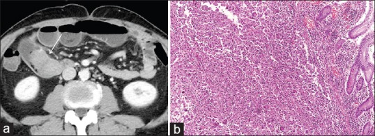
Figure 1.

(a) Abdominal computed tomography shows a 6-cm enhancing mass in the small bowel that is obstructing the entire lumen (arrow). There is luminal dilatation of the distal loop of the small bowel. (b) High-power photomicrograph (original magnification, ×100; H and E stain) of the specimen obtained by small bowel resection shows a multinucleated giant-cell carcinoma. H and E: hematoxylin and eosin
