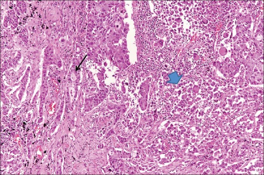
Figure 4.

High-power photomicrograph (original magnification, ×200; H and E stain) of a surgical specimen obtained at lobectomy of the lung mass shows the adenocarcinoma component on the left (arrow) and giant-cell carcinoma on the right, central direction (arrowhead) being sharply separated from each other. H and E: hematoxylin and eosin
