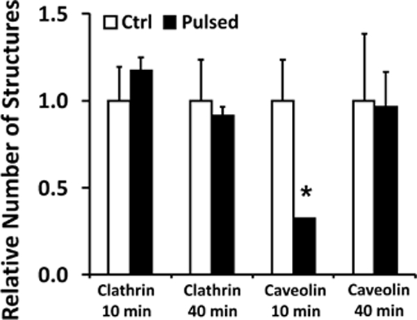
Figure 3. Effects of cell treatment with electric pulses on numbers of small vesicles.

COS7 cells in the buffer containing non-labeled pDNA were treated with five electric pulses (400 V/cm, 5 msec, and 1 Hz). Cells in the control groups were prepared with the same procedures except that the electric pulses were not delivered. At 10 or 40 min post treatment, membranous structures in cells were post-stained with uranyl acetate and lead citrate to enhance their contrast under the electron microscope (see the Materials and Methods section), but no immunostaining was performed for these cells. Subcellular structures of pits and vesicles (50 – 100 nm in diameter) were grouped together. The structures with smooth and rough membranes were considered to be clathrin- and caveolin-coated, respectively. The total numbers of the structures were counted in 10 different cells (i.e., one section per cell); and the data in the pulsed groups were normalized by the corresponding controls. Bars and error bars represent the mean and the standard error of the mean, respectively. *p < 0.05, Ctrl vs Pulsed.
