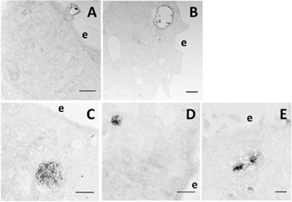
Figure 4. Representative electron micrographs of pDNA-positive structures.

Morphological criteria were developed in the study for the identification of subcellular structures associated with pDNA. Some morphological features are shown in Figs. 1 and 2, respectively. Others are shown in this figure. (A) A membrane protrusion/ruffle with a closure is associated with the EDS in a COS7 cell, indicating that macropinocytosis is involved in pDNA uptake. Bar = 500 nm. (B) An early endosome-like structure with an electron-translucent lumen contains a few internal vesicles that are positive for the EDS in an HCT116 cell. Bar = 500 nm. (C) A large and (D) a relatively smaller late endosome/lysosome-like structures are positive for the EDS in COS7 cells. Bar = 1 μm. (E) A few electron dense aggregates can be seen in an HT29 cell; and each aggregate is surrounded by multiple vesicles. Bar = 200 nm. e, extracellular space.
