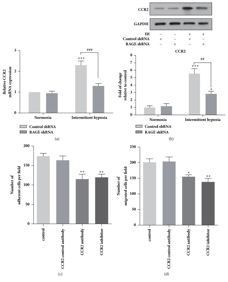
Figure 3.
Intermittent hypoxia promoted CCR2 expression via RAGE, which mediated chemotaxis and adhesion of THP-1 cells. Expression levels of CCR2 in THP-1 cells exposed to normoxia and intermittent hypoxia determined by qRT-PCR (a) and western blotting (b). The effects of CCR2 neutralizing antibody (10 μg/m) and CCR2 inhibitor (10 nM) on (c) the adhesion of THP-1 monocytes to HUVECs and the cell transwell migration (d). Data were represented as mean + SEM. ∗ represents significant difference compared with control group under normoxia condition; ∗p<.05, ∗∗/##p<.001, ∗∗∗/###p<.001.

