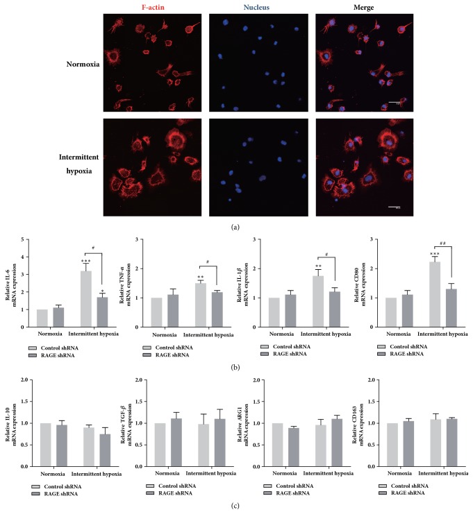
Figure 4.
Intermittent hypoxia polarizes macrophages to an M1 phenotype, which was mediated by RAGE. (a) Morphology of THP-1 macrophage cultured in normoxia or IH. F-actin cytoskeleton of the cells were labelled with phalloidin (red) and nuclei stained with DAPI (blue). Scale bar: 20 μm. (b) mRNA expression levels of M1 expression markers. (c) mRNA expression levels of M2 expression markers. ∗ represents significant difference compared with control group under normoxia condition; ∗p<.05, ##p<.001, ∗∗∗/###p<.001.

