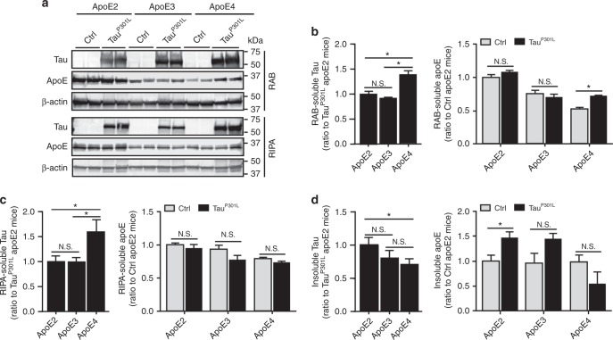
Fig. 3.
Increased insolubility of tau and apoE in TauP301L-apoE2 mice. The cortical brain tissues from control mice (Ctrl, AAV-GFP-injected) and TauP301L-apoE2, -apoE3, and -apoE4 mice at 6 months of age were sequentially extracted by RAB, RIPA, and FA buffer. Soluble tau (HT7 detection) and apoE in RAB (a, b) and RIPA fractions (a, c) were examined by western blotting (n = 6 mice per group, mixed gender). Results were normalized to β-actin levels. Insoluble tau and apoE in FA fraction was detected by ELISA (d n = 6 mice per group, mixed gender). Data are expressed as mean ± SEM relative to apoE2-TR mice. Mann-Whitney tests followed by Bonferroni correction for multiple comparisons were used. *P < 0.0167; N.S. not significant

