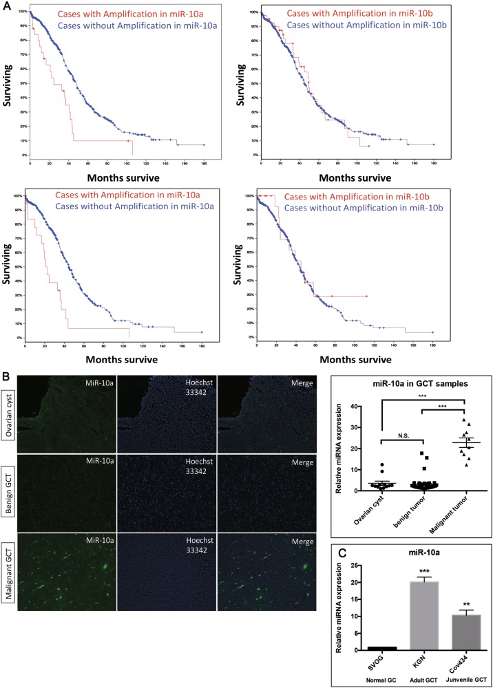
Fig. 1. MiR-10 in ovarian cancer patients and granulosa cells tumor and cell lines.
a Above and below are two independent groups of ovarian cancer patients. Lower survival rate of ovarian cancer patients for those cases with amplification of miR-10a, but not miR-10b. b Fluorescence in situ hybridization (FISH) studies detected a stronger staining in malignant GCT tissues than either begin GCT tissues or ovarian cysts. c The level of miR-10a increases in two GCT cell lines KGN and Cov434. Each bar in the figure represents the mean ± SEM of triplicates. ** P < 0.01 and *** P < 0.0001

