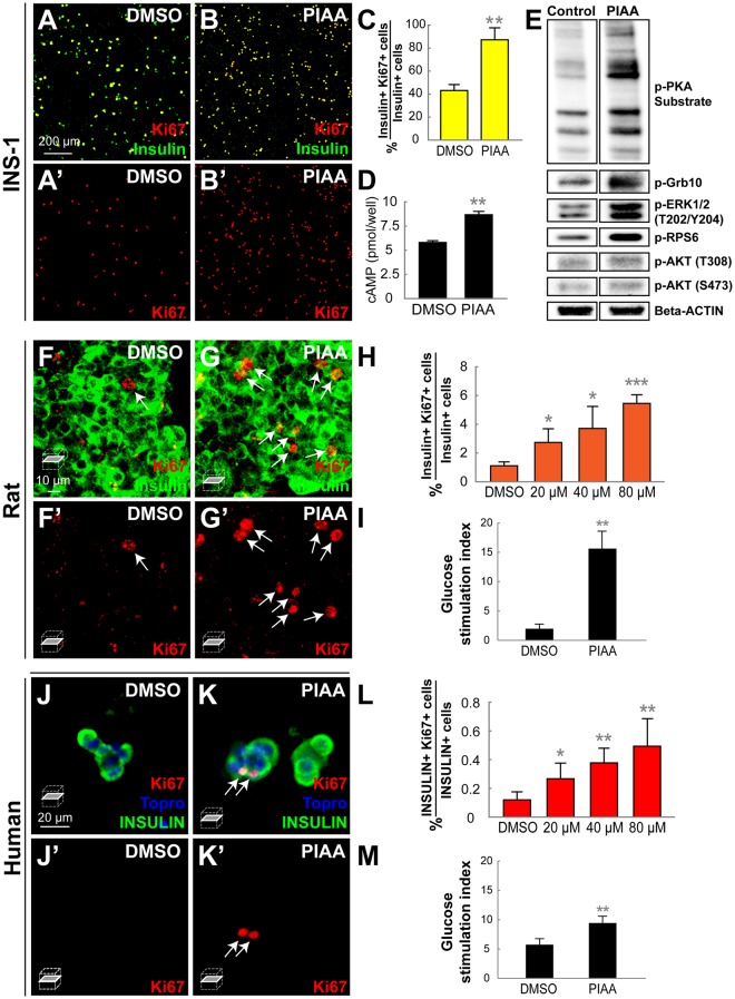
Figure 7.
PIAA induces proliferation of rat and human β-cells. (A-B’) Confocal images of rat INS-1 β-cells treated with DMSO (A-A’) and PIAA (B-B’), respectively, stained for Ki67 (red) and Insulin (green). (C) The percentage (mean ± SD) of Ki67 and Insulin-double positive cells (in A-B’; 43.1 ± 5.2% (DMSO) and 87.3 ± 10.4% (PIAA)). (D) Quantification of cAMP levels (mean ± SD) (5.8 ± 0.2 pmol/well (DMSO) and 8.7 ± 0.4 pmol/well (PIAA)). (E) representative Western blot showing increased phosphorylation of PKA substrate and ERK1/2 in PIAA-treated INS-1-derived 832/13 β-cells. Note that PIAA-treatment augmented phosphorylation of mTOR targets RPS6 and Grb10 but did not trigger phosphorylation of AKTT308 and AKTS473. (F-G’) Confocal single-plane images of whole rat islets treated with DMSO (F-F’) and PIAA (G-G’), respectively, stained for Ki67 (red, white arrows) and Insulin (green). (H) The percentage (mean ± SD) of Ki67 and Insulin-double positive cells in whole rat islets increased in a dose-dependent manner with treatment of PIAA (1.1 ± 0.3% (DMSO), 2.7 ± 0.9% (20 μM), 3.7 ± 1.5% (40 μM), and 5.5 ± 0.6% (80 μM)). n = 5 replicates per condition. (I) Glucose stimulation indices of rat islets treated with DMSO or PIAA (300 islet equivalents per column, triplicate). (J-K’) Confocal single-plane images of human islets treated with DMSO (J-J’) and PIAA (K-K’), respectively, stained for Ki67 (red, white arrows), Topro (blue), and INSULIN (green). (L) The percentage (mean ± SD) of Ki67 and Insulin-double positive cells in human islets increased in a dose-dependent manner with treatment of PIAA (0.1 ± 0.0% (DMSO), 0.3 ± 0.1% (20 μM), 0.4 ± 0.1% (40 μM), and 0.5 ± 0.2% (80 μM)). n = 5 replicates per condition from 3 cadaveric donors. (M) Glucose stimulation indices of human islets treated with DMSO or PIAA (300 islet equivalents per column, triplicate). *P < 0.05; **P < 0.01; ***P < 0.001.

