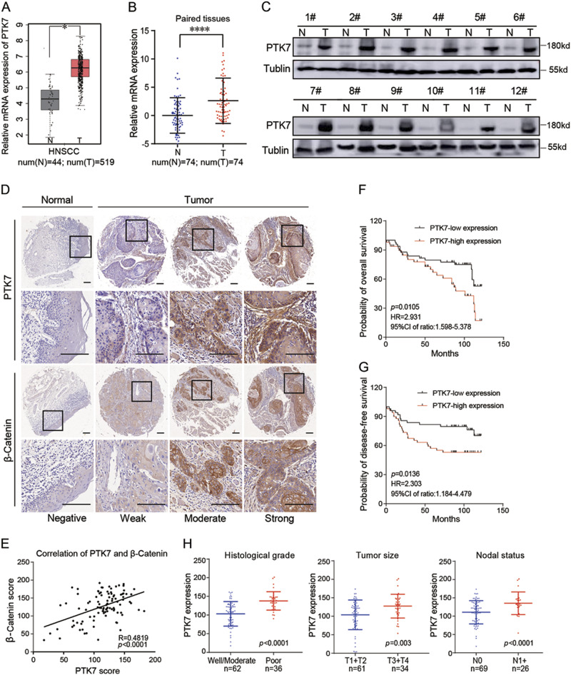
Fig. 1. Increased PTK7 expression is correlated with the Wnt/β-Catenin pathway and aggressive clinicopathologic features and predicts a poor prognosis in HNSCC.
a Relative PTK7 mRNA expression levels in normal tissues and HNSCC tissues from TCGA database are shown. *p < 0.05. b Relative PTK7 mRNA levels in 74 paired adjacent normal tissues and HNSCC tissues were evaluated by RT-PCR. ****p < 0.0001, based on the paired t-test. c PTK7 protein expression levels in 12 paired adjacent normal tissues and HNSCC tissues were determined by western blot. Tubulin was used as a loading control. d IHC analysis of PTK7 and β-Catenin expression levels in tissue microarrays containing 10 normal tissues and 98 HNSCC tissues. Images of negative, weak, moderate, and strong PTK7 and β-Catenin staining are shown. Scale bar: 20 μm. e The correlation of PTK7 and β-Catenin expression was analyzed in tissue microarrays containing 10 normal tissues and 98 HNSCC tissues (R = 0.4819, p < 0.001). f OS was significantly different between the low and high PTK7 expression groups in HNSCC (p = 0.0105, HR = 2.931, 95% CI of ratio: 1.598–5.378). g DFS was significantly different between the low and high PTK7 expression groups in HNSCC (p = 0.0136, HR = 2.303, 95% CI of ratio: 1.184–4.479). h PTK7 protein expression levels were significantly associated with historical grade, tumor size, and nodal status

