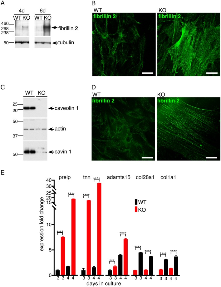
Fig 3. Fibrillin2 protein levels are increased in CAV1-/- MEFS and CAV1 KO NIH-3T3 cells.
A. Western blots with anti-fibrillin-2 or anti-tubulin antibodies of lysates from CAV1-/- MEFs (KO) and congenic WT controls harvested at the indicated number of days after plating. B. Indirect immunofluorescence with anti-fibrillin-2 antibodies labelling either CAV1-/- MEFs (KO) or congenic WT controls fixed and stained four days after plating. Bars 10μ. C. Western blots to demonstrate absence of caveolin 1 in CRISPR-generated CAV1 knockout NIH3T3 cells. Absence of caveolin1 results in reduced expression of cavin1. D. Indirect immunofluorescence with anti-fibrillin-2 antibodies labelling either control NIH3T3 cells (WT) or CAV1 null NIH3T3 cells (KO). Bars 10μ. E. Quantitative PCR was used to measure the levels of the transcripts shown in mRNAs purified from CAV1 KO NIH-3T3 cells and WT controls. Expression fold change is relative to the WT, 3 days culture sample. The cells were grown in culture for the number of days shown. Bars are SD, N = 4 experimental repeats. P values were determined using a T-test.

