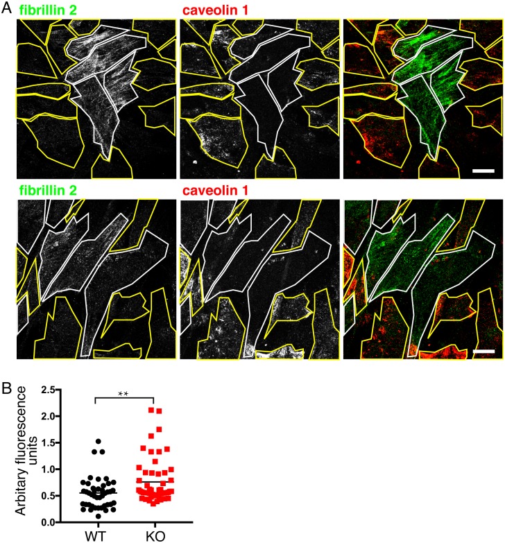
Fig 4. Increased fibrillin 2 expression in CAV1-/- MEFS is maintained during co-culture with WT MEFs.
A. Indirect immunofluorescence with anti-fibrillin-2 and anti-caveolin-1 antibodies labelling CAV1-/- MEFs (KO) and congenic WT controls plated as a mixed culture for 10 days. Two representative fields of cells are shown, KO cells identified by absence of caveolin 1 signal are outlined in white, WT cells are outlined in yellow. Bars 10μ. B. Quantification of fibrillin 2 expression in WT and CAV1-/- MEFs plated as mixed cultures as in A, expressed in arbitrary fluorescence units from the mean fluorescence intensity of individual cell areas after background subtraction. P value was determined using a T-test.

