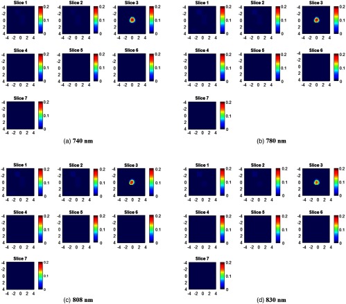
Fig. 3.
Target absorption maps (a) 730 nm, (b) 785 nm, (c) 808 nm, and (d) 830 nm of a SHC phantom located at 1.0 cm depth (target top position). For each absorption map, seven slices from 0.5 to 3.5 cm depth with 0.5-cm increment have reconstructed. The spatial dimensions of each slice are . Color bar is the absorption coefficient in the unit of .

