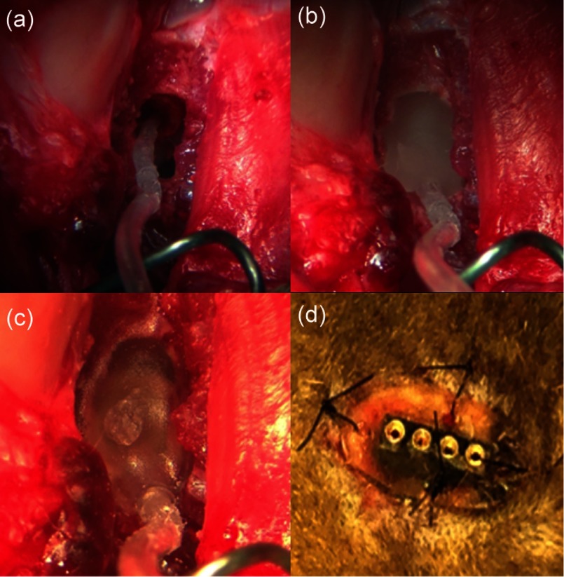
Fig. 4.
The implantation of the optrode into a cat cochlea. (a) The optrode was inserted into a cat cochlea through the cochleostomy, (b) the optrode was fixed to bulla with dental acrylic, (c) the second layer of dental acrylic, and (d) the transcutaneous connector was secured onto the lower neck skin.

