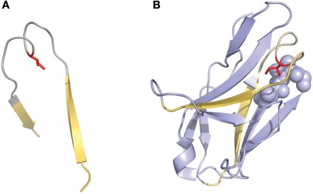
Figure 3.

Representation of the “two-span-bridge” conformation of CDRL1 present in the 2fb4 antibody structure. The two-span-bridge is represented in gray and the bordering strands are shown in yellow (A, B). (A) Shows the isolated loop with an Ile side chain (colored red) pointing inwards the loop structure. (B) The complete domain showing how the Ile penetrates deeply into the core of the light chain variable domain making hydrophobic interactions with neighboring residues organized in a pocket (purple spheres).
