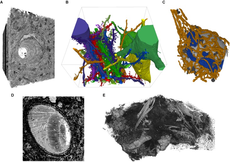
FIGURE 5.

Example screenshots of the 3D viewer. (A) Capillary running through cortex, rendered from EM image stack. (B) Segmented spiny dendrites and cell bodies (fully manual segmentation). (C) Organelles in a neuron soma; mitochondria in orange, Golgi apparatus in blue, lysosomes dark gray, nucleus light gray (fully manual segmentation). (D) Erythrocyte in a capillary in LGN rendered from an EM image stack. (E) Micro-CT scan of a fossil specimen of Paleothyris acadiana (Museum of Comparative Zoology, Harvard). Data in A–C from (Kasthuri et al., 2015); D from (Morgan et al., 2016); E with kind permission of S. Pierce, Museum of Comparative Zoology, Harvard.
