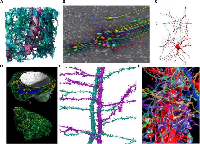
FIGURE 6.
Examples of 3D models segmented in VAST and rendered in Autodesk 3ds Max. (A) Spiny dendrite (red) and axons (green) in a 10 μm × 10 μm × 6 μm cube of mouse cortex. At a voxel size of 6 nm × 6 nm × 30 nm, even the finest neural processes are segmentable. (B) Neurons traced in a low-resolution EM stack of mouse cortex, shown in situ above one EM section, with apical dendrites running in a bundle toward the pia (to the right in the image). Field of view roughly 510 × 340 micrometers. (C) Putative basket cell in rat cortex, traced semi-automatically with trans-layer masking in a ∼100 μm × 100 μm × 200 μm tissue volume. Tracing this cell took ∼15 h for a single expert. (D) Organelles in a neuron cell body (same cell as Figure 5C) shown from two directions. Nucleus white, with pores visible; endoplasmic reticulum green, mitochondria yellow, Golgi apparatus blue. (E) Two spiny apical dendrites with side branches in rat cortex. Synapses shown in yellow. (F) Spiny dendrite in red, with transparent axons making synapses on it. Neurotransmitter vesicles (white) were exported using particle clouds to generate spherical vesicles. All images are based on EM data from the lab of Jeff Lichtman, Harvard. All segmentations except C were done fully manually. A,B,D,F used data published with (Kasthuri et al., 2015).

