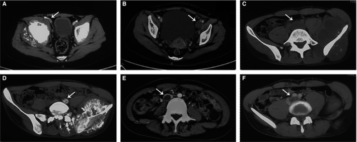
Figure 1.

Manifestations of venous tumor thrombus (VTT) on plain and enhanced CT scan. A, A VTT located at the right external iliac vein (arrow) showed unchanged caliber of the vessel compared with the contralateral side. B, A VTT located at the left external iliac vein (arrow) showed enlarged caliber of the vessel compared with the contralateral side. C, A VTT located at the left common iliac vein (arrow) showed low density compared with the muscles on plain CT scan. D, A VTT located at the left common iliac vein (arrow) showed apparent calcification within the vascular lumen on plain CT scan. E, A VTT located at the inferior vena cava (arrow) showed filling defect within the vascular lumen on contrast enhancement. F, A VTT located at the left common iliac vein (arrow) showed streak‐like enhancement within the filling defect on contrast enhancement
