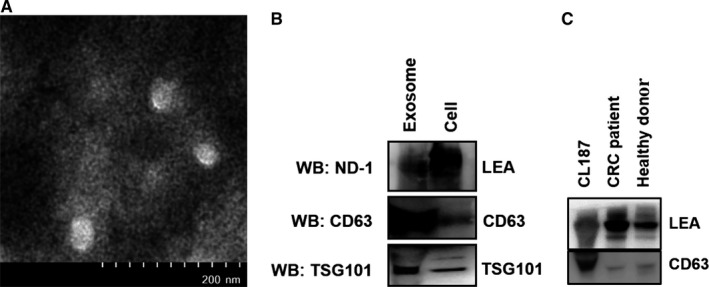
Figure 6.

Characterization of exosomes derived from CL187 cells and CRC patient plasma. A, Representative electron microscope images of exosomes isolated from CL187 cells. B, Western blotting analysis of LEA and exosome‐marker expression in exosomes from CL187 cells using ND‐1, anti‐TSG101, and anti‐CD63, respectively. C, Representative analysis of LEA expression in exosomes from CRC patient and healthy donor plasma by western blotting analysis using ND‐1
