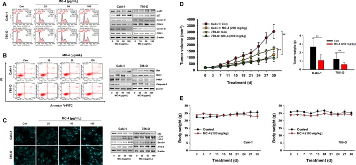
Figure 2.

Anticancer effects of MC‐4 in human RCC cells. A, Effects of MC‐4 on the cell cycle. Caki‐1 and 786‐O cells were treated with MC‐4 (25, 50, or 100 μg/mL) for 24 h. After incubation, the cells were stained with propidium iodide (PI) and then analyzed using a flow cytometer (left panel). The expression levels of cell cycle‐regulated proteins were measured by Western blot analysis (right panel). B, Effects of MC‐4 on apoptosis. Scatter plots present the percentage of viable, early apoptotic, late apoptotic, and necrotic cells in untreated (control) and treated cells (left panel). The cells were treated with MC‐4 (25, 50, or 100 μg/mL) for 24 h. The expression levels of apoptosis‐related proteins were measured by Western blotting (right panel). C, Effects of MC‐4 on autophagy. Monodansylcadaverine (MDC) staining shows that autophagy was activated in Caki‐1 and 786‐O cells after treatment with MC‐4. Magnification ×400 (left panels). The expression levels of autophagy‐related proteins in RCC cells were measured after MC‐4 treatment. Western blot analysis was performed using p62, beclin‐1, LC3‐I/II, and ATG5 antibodies. Equal loading and transfer were verified by reprobing the membranes with β‐actin antibody (right panels). D, Effects of MC‐4 on tumor growth in nude mice inoculated with Caki‐1 or 786‐O cells. During the 30‐d treatment, tumor volumes were estimated using measurements taken with calipers (mm3). The histogram data shown are average tumor volumes and tumor weights (mean ± SEM, n = 5). **P < 0.01 vs vehicle control. E, Effects of MC‐4 on body weight changes in nude mice inoculated with Caki‐1 (left) and 786‐O (right) cells
