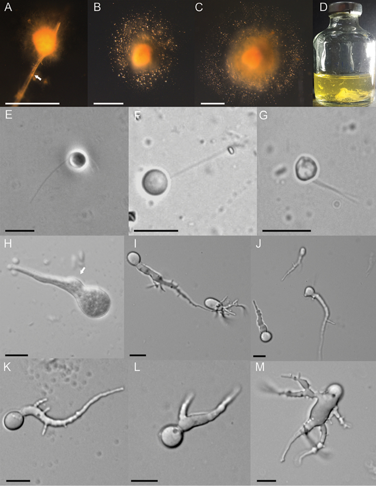
Figure 1.
Macroscopic and microscopic features of Liebetanzomycespolymorphus. Colony morphology on agar roll tubes (A–C), showing the development of a colony attached (A) to a straw particle (arrowed), dense growth in the centre surrounded by numerous sporangia and zoospores (B–C) causing expansion of colony size. Growth in liquid medium showing a biofilm-like growth (D). Zoospores are spherical and Uniflagellate (E–F) or biflagellate (G). Germinating zoospore (H) showing a zoospore cyst (arrowed), presence and absence of sporangiophore indicating the endogenous and exogenous type of sporangial development (I) and different shapes of sporangia (I, J). Early stages of thallus development showing a single (K), bifurcated (L) and multifurcated (M) rhizoidal system. Scale bar: 1 mm (A–C); 10 µM (E–M).

