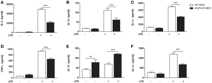
Figure 4.
AQP9-KO DCs cytokine profile following LPS exposure. BMDCs from WT and Aqp9-KO mice were exposed to LPS for 24 h. (A–F) Cytokine (IL-6, IL-12, IL-1α, TNFα, IL-10, IL-1β) concentrations were determined in the supernatant by ELISA. Bars represent mean ± SEM of 3 independent experiments. **P < 0.01, ***P < 0.001.

