
Figure 1.
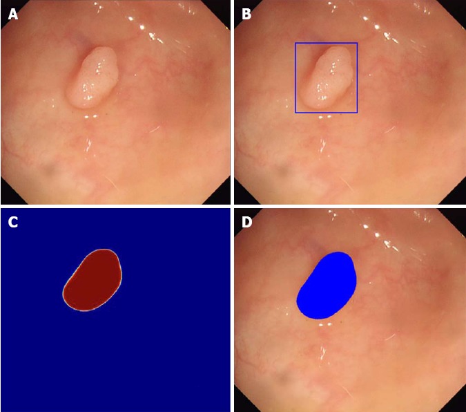
Automatic polyp detection by Wang et al[40]. A: Original image obtained during colonoscopy; B: Automatic detection by box method; C: Probability map whereby red indicates high probability of polyp and blue indicates low probability of polyp; D: Automatic detection by paint method whereby blue coloring indicates location of polyp.