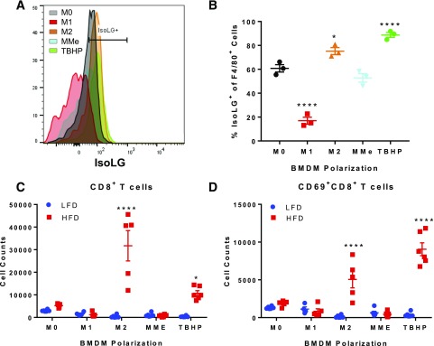
Figure 8.
M2 polarization increases IsoLGs levels and promotes activation of CD8+ T cells. BMDMs were polarized to M0, M1, M2, or MMe or treated with TBHP. After polarization, BMDMs were cocultured with isolated T cells from the spleen of mice fed an LFD or HFD for 3 days. IsoLG content was quantified in polarized BMDMs by flow cytometry. A and B: Flow analysis (A) and quantification (B) of IsoLG+ F4/80+ cells (one-way ANOVA). Data are mean ± SEM (n = 3 wells/group). T cells were collected from cocultures, and flow analysis was performed to quantify CD8+ T cells (C) and CD8+CD69+ T cells (D) (two-way ANOVA). Data are mean ± SEM (n = 5 mice/LFD or HFD group) and mean ± SEM (n = 5 wells/group). *P ≤ 0.05, ****P ≤ 0.0001.

