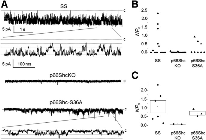
Figure 5.
Knockout of p66Shc inhibits KATP channels. A: Representative cell-attached patch-clamp recording of vascular SMC isolated from wild-type, p66ShcKO, and p66Shc-S36A mutant rats. Membrane potential (Vp) = 0 mV. Scales are the same for all three recordings. Activity is also shown at an expanded scale. The closed state (c) is upward. B: Scatter plot demonstrate individual channel activity (NPo) for KATP channels recorded in cell-attached patches from SS, p66ShcKO, and p66Shc-S36A SMC cells at a 0 mV holding potential. C: NPo for experiments with observed channel activity (shown in B). Data are the mean ± SE.

