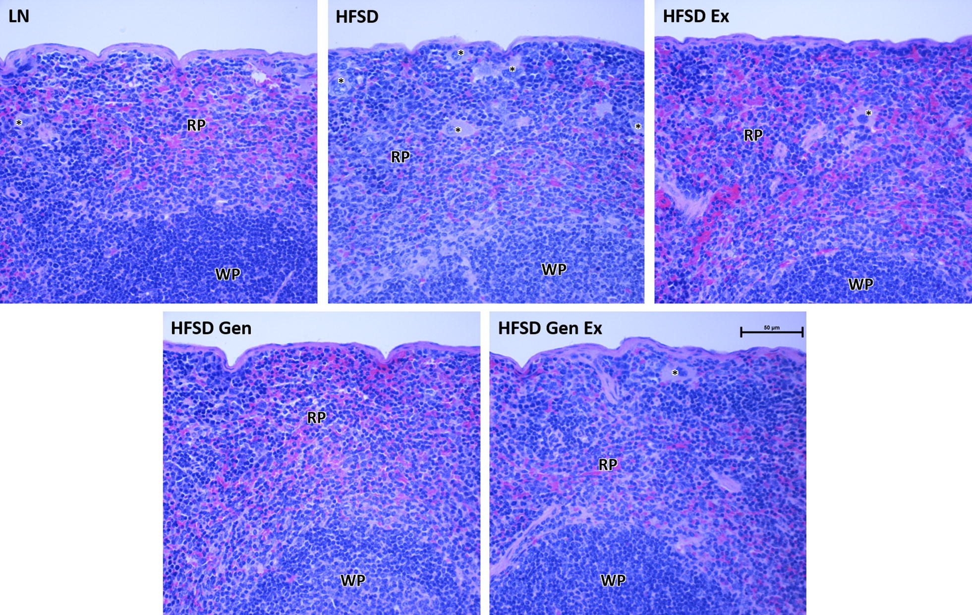
Fig. 3.

Representative histological sections of the spleen for each treatment group. Note the high cellularity of the control HFSD-fed mice, which is not present to the same extent in the HFSD treated with exercise and/or genistein. Mice fed HFSD also have numerous macrophages in the red pulp (asterisks) in comparison to the other treatment groups. Splenic morphological appearance did not differ by sex. LN, lean mice fed standard diet (n = 10 females, 8 males); HFSD, high-fat, high-sugar diet (n = 9 females, 9 males); Ex, exercise (n = 9 females, 10 males); Gen, genistein (n = 8 females, 8 males); GenEx, genistein and exercise (n = 10 females, 8 males). RP, red pulp; WP, white pulp. Histological analysis was conducted on all 100 mice. H&E stain. Scale bar 50 μm
