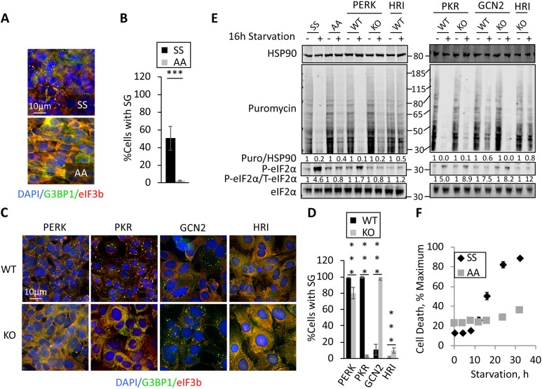
Fig. 4.
Role of eIF2α kinases in translation repression and stSG induction during chronic nutrient starvation. (A) Wild-type or S51A eIF2α mutant MEFs were stressed for 12 h prior to fixation and staining of stSGs with antibodies against G3BP1 (green) or eIF3b (red). (B) Quantification of the results shown in A. Statistical analysis was performed with a Chi-test (***P<0.001). (C) Wild-type or eIF2α kinase KO MEFs indicated were stressed as in A followed by fixation and staining. (D) Quantification of the results shown in C. Statistical analysis was performed with a Chi-test (***P<0.001). (E) MEFs were starved and lysates were prepared for western blotting for the puromycin-labeled polypeptides to assess ongoing translation, or the indicated proteins. Molecular weight standards are indicated. Quantification of western blots was performed using image studio for Li-Cor to avoid saturation effects, and ratios of total to phosphoproteins are indicated. (F) Wild-type and S51A eIF2α mutant MEFs were starved in the presence of ethidium homodimer-1, as described above. Ethidium fluorescence was normalized to 0 h and plotted against time. Results from all panels are representative of a minimum of three experimental replicates, and data are presented as mean±s.d.

