Fig. 5.
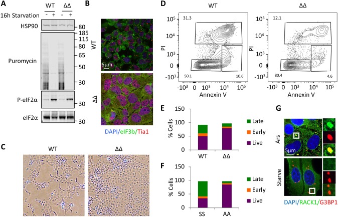
Chronic nutrient starvation induces pro-death stSG. (A) Wild-type and ΔΔ U2OS cells were starved for 16 h, followed by western blotting for puromycin or the indicated proteins. Molecular weight standards are indicated. (B) Cells treated as in A were stained for stSGs using eIF3b (green) and Tia1 (red) after 16 h starvation. (C) Phase-contrast images taken with a 10× objective, showing the appearance of the respective monolayers for wild-type and ΔΔ U2OS cells. (D) Flow cytometry was performed on fed and starved U2OS cells (Fig. S4) stained with annexin V and propidium iodide to monitor the proportion of cells undergoing cell death. (E) Graphical illustration of flow cytometry results in U2OS cells. The y-axis is the percentage of cells in each category, with the total equaling 100%. (F) Wild-type and S51A eIF2α mutant MEFs were subjected to the same analysis as in D and are represented as in E. (G) U2OS cells were stressed with arsenite or starved for 16 h, as indicated, before fixation and staining for RACK1 (green) and G3BP1 (red). Results from all panels are representative of a minimum of three experimental replicates.

