
Fig. 3.
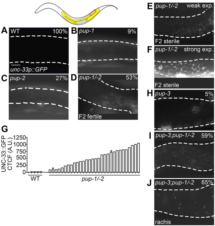
Expression of a pan-neuronal reporter in pup mutant germlines. Diagram shows the mid-late pachytene region of the germline represented in A-F,H-J. (A) unc33p::gfp expression was not detected in otherwise wild-type germ cells. Relatively low abundance unc33p::gfp expression was detected in (B) 9% of pup-1(0) F2 germlines and (C) 27% of pup-2(0) F2 germlines. For pup-1, GFP was limited to localized sets of a few germ cells; for pup-2, GFP extended more broadly. In all cases, GFP was observed in cortical cytoplasm and not in the rachis. unc-33p::gfp expression was detected in (D) 53% of fertile and (E,F) 100% of sterile pup-1/-2 F2 (M–Z–) germlines. (G) Quantification of GFP expression in control (n=5) and pup-1/-2(0) mutant (n=30) germlines. Each bar represents a single germline, and asterisk indicates the individuals pictured in E and F, respectively (see Materials and Methods). CTCF, corrected total cell fluorescence. (H) Very low cortical unc-33p::gfp expression was observed in 95% of pup-3(0) germlines; localized regions of stronger expression were observed in 5% of pup-3(0) germlines, as pictured. (I) Cortical unc-33p::gfp expression was observed in 59% of pup-3;pup-1/-2 F2 (M−Z–) germlines. (J) GFP puncta were also observed in the cytoplasmic core (rachis) of the germline in 65% of pup-3;pup-1/-2 F2 (M−Z–) gonad arms. Scale bars: 16 µm.