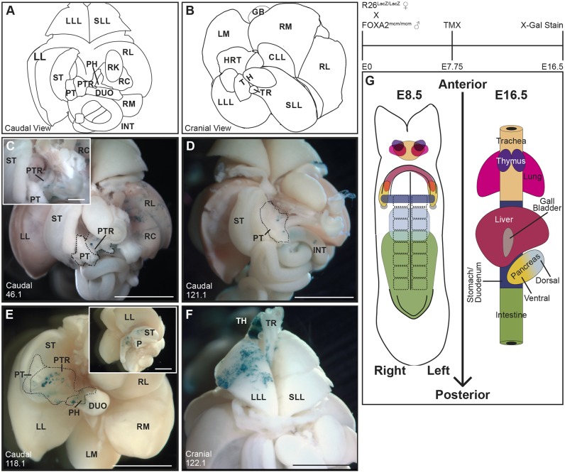
Fig. 1.
Lineage tracing reveals individual multi-organ progenitors in the E8.5 DE. Three mg kg−1 TMX was maternally administered at E7.75 to Foxa2mcm/+; R26R−/+ embryos and X-gal staining of dissected viscera performed at E16.5. (A,B) Illustrated and annotated caudal (A) and cranial (B) views of the dissected viscera are provided for reference. (C) A caudal view of embryo 46.1 reveals lacZ-expressing descendants in the right lateral (RL) and right medial (RM; not seen in this view) liver lobes, as well as in the pancreatic trunk (PTR; n=3 embryos with this pattern). (D) A caudal view of the viscera dissected from embryo 121.1 reveals blue lacZ-expressing cells in the pancreatic tail (PT) and intestine (INT; n=2 with this pattern). (E) A caudal view of the viscera of embryo 118.1 reveals lacZ-positive cell descendants in the stomach (ST) as well as in the PTR and pancreatic head (PH). The left/caudal view of this viscera in the inset reveals additional lacZ-positive descendants in the ST and duodenum (DUO; n=3 with this pattern). (F) A cranial view of embryo 122.1 demonstrates lacZ-expressing clonal descendants in the left lung lobe (LLL), left thymus (TH) and in the trachea (TR). (G) A fate map of the E8.5 DE, produced by combining previously published fate mapping with the novel progenitor relationships reported herein. The fate map is drawn on a linearized 8S embryo. The midline placed notochord, paired somites, and anterior and posterior intestinal portal are drawn on the embryo as landmarks. The shapes indicate organ progenitor domains and the color represents individual organs as identified on the simplified E16.5 gut tube. Progenitor domains that overlap represents endoderm that will contribute to each of those organs. CLL, cardiac lung lobe; GB, gall bladder; HRT, heart; LL, left lateral liver lobe; LM, left medial liver lobe; RC, right caudate liver lobe; RK, right kidney; SLL, superior lung lobe. Scale bars: 1 mm.

