Fig. 2.
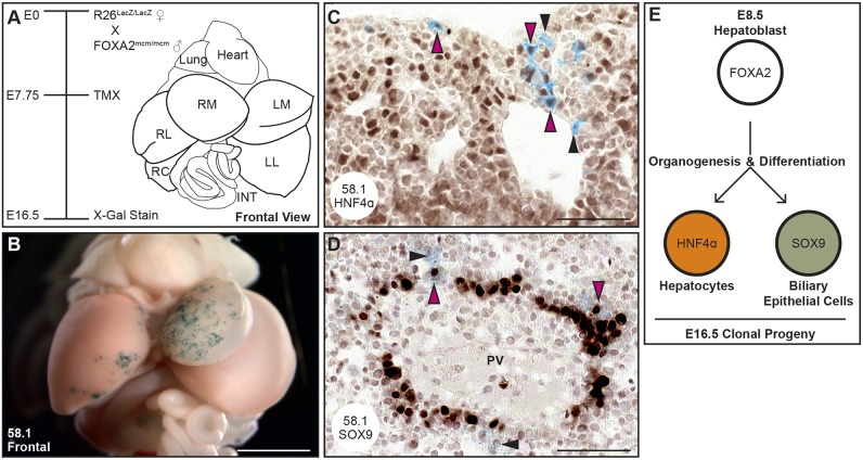
Individual hepatoblasts contribute hepatocytes and BECs to each E16.5 liver. Embryos were maternally administered 3 mg kg−1 TMX at E7.75 and then dissected and X-gal stained at E16.5. (A) An annotated illustration of a frontal view of dissected viscera at E16.5. (B) A frontal image of the viscera of embryo 58.1 reveals widespread lacZ expression in the left medial liver lobe (LM) and more restricted expression on the left edge of the right medial liver lobe (RM). (C,D) Immunohistochemistry, performed on sections from the embryo in B, reveals the colocalization of lacZ-expressing clonal descendants (blue) with the hepatocyte marker HNF4α (C, pink arrowheads) and the biliary epithelial cell marker SOX9 (D, pink arrowheads). The black arrowheads indicate lacZ-expressing descendants that are negative for the indicated immunohistochemistry marker. (E) Schematic illustrating that each clonally labeled E8.5 hepatoblast contributes to the hepatocyte and biliary epithelial cell lineage by E16.5 (n=22/22; χ2, ***P=9.11−4). Scale bars: 1 mm in B; 50 µm in C,D. INT, intestine; LL, left lateral liver lobe; RC, right caudate liver lobe; RL, right lateral liver lobe.

