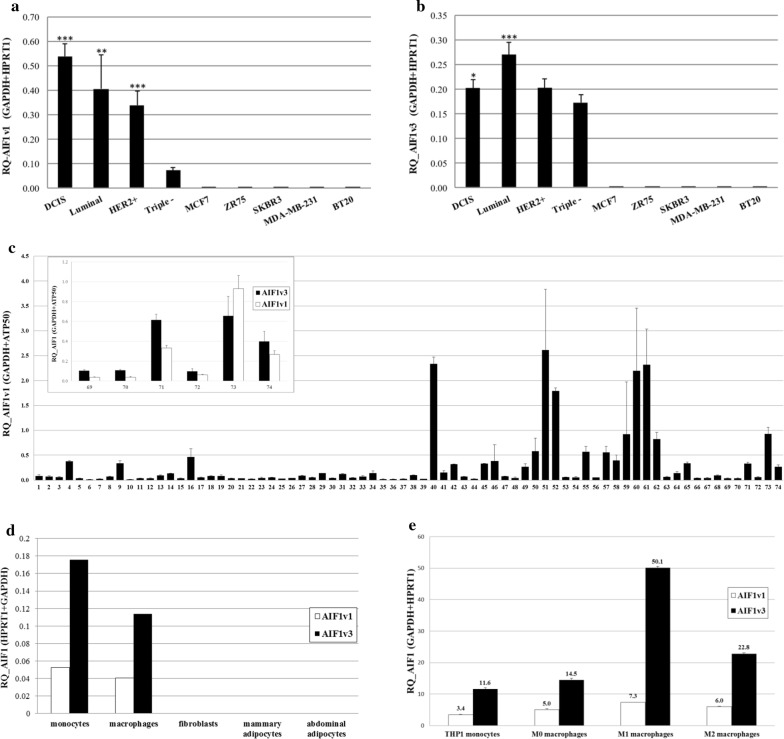
Fig. 3.
Characterization of AIF1 isoforms in breast tumors and tumor microenvironment. Expression of a AIF1v1 and b AIF1v3 in breast tumors of varying severity (DCIS, luminal A/B (ER+ and/or PR+), HER2+ (ER−/PR−/HER2+), triple negative (ER−/PR−/HER2−) and human BC cell lines (MCF7, ZR75, SKBR3, MDA-MB-231, BT20). c AIF1v1 in breast adipose tissue (top panel: comparison of AIF1v1 and AIF1v3 expression in six samples of breast adipose tissue). d AIF1 isoforms in various cell types of the breast tumor microenvironment (monocytes, macrophages, fibroblasts and adipocytes). e AIF1 isoforms in THP-1 monocytes and differentiated macrophages (M1, M2). Data shown as mean ± SD; Each subtype is compared to triple negative with ***p < 0.01, **p < 0.05, *0.05 < p < 0.1

