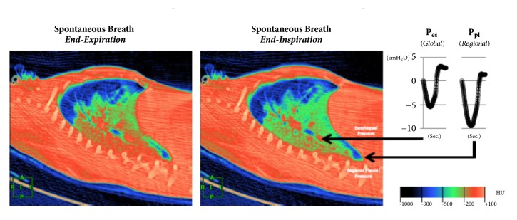
Figure 2.
Dynamic CT scan in end-expiration (left panel) demonstrates that the aerated lung (blue) is nondependent, while the dependent lung is densely atelectatic (red). At end-inspiration during a spontaneous breath (mid panel), there is little change in the nondependent aerated lung (blue); the dependent lung, previously densely atelectatic (red) is now partially aerated (green/red). The inspiratory pleural pressure traces (right panel), measured at the arrow tips, show the negative deflections (“swings”) in regional Ppl and global Pes during inspiration. However, the “swing” in regional Ppl is greater (x2) than the “swing” in Pes, indicating that diaphragm contraction results in greater distending pressure applied to the regional lung near the diaphragm compared with the pressure transmitted to the remainder of the lung (i.e., Pes). Ppl: pleural pressure; Pes: esophageal pressure; HU: Hounsfield Units, with authors permission [15].

