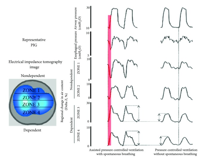
Figure 4.
Electrical impedance tomography (EIT) waveforms in experimental lung injury, spontaneous versus ventilator breaths. In an anesthetized pig model of acute lung injury assist pressure-controlled ventilation (IP, 15 cm H2O; f, 25 min21; PEEP=13 cm H2O; triggering threshold, 22 cm H2O) was used. The EIT image was divided into four zones, each covering 25% of the ventrodorsal diameter (zones 1–4). During controlled ventilation (under muscle paralysis), simultaneous inflation of each of the different lung regions was observed, although at different inflation rates. In contrast, when spontaneous efforts were present, two observations were noted. First, in the initial stages of the breath, spontaneous efforts caused inflation of dependent lung regions (red in zones 3 and 4), which was greater with controlled breaths. Second, the early inflation in the dependent region was accompanied by concomitant (transient) deflation of nondependent region (red in zone 1), indicating movement of gas from nondependent to dependent lung regions, because this was not associated with alterations in tidal volume it indicates a pendelluft phenomenon. This finding was always present during spontaneous breathing efforts in all animals with experimental lung injury: f = respiratory frequency; IP: inspiratory pressure; PEEP: positive end-expiratory pressure, with authors permission [16].

