Figure 1.

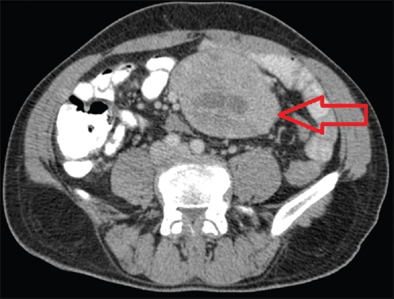
Contrast-enhanced computed tomography showing a soft tissue mass (red arrow) 7.2 × 8.4 cm with necrotic areas abutting small bowel loops

Contrast-enhanced computed tomography showing a soft tissue mass (red arrow) 7.2 × 8.4 cm with necrotic areas abutting small bowel loops