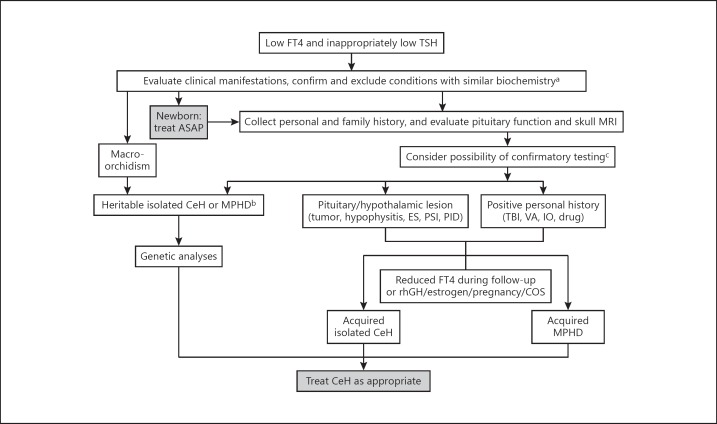
Fig. 1.
Flow chart for the diagnosis and management of CeH. MRI, magnetic resonance imaging; CeH, central hypothyroidism; MPHD, multiple pituitary hormone defect; ES, empty sella; PSI, pituitary stalk interruption; PID, pituitary infiltrative disease; TBI, traumatic brain injury; VA, vascular accident; IO, iron overload or hemochromatosis; rhGH, recombinant human growth hormone; COS, controlled ovarian stimulation. a Confirm low FT4 and inappropriately low TSH, and exclude conditions reported in Table 3. b See Table 1 for details. c See Table 4 for details.

