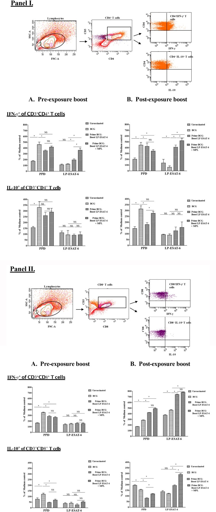
Figure 5.
Intracellular IFN-γ and IL-10 are differentially expressed in antigen-specific CD4+ (I) and CD8+ (II) T cells in BCG-primed mice boosted with LP-ESAT-6 subunit vaccine in pre-exposure or post-exposure schedule. Female BALB/c mice (n = 5/group) were immunized according to the pre-exposure or post-exposure schedule shown in Figure 1. Four weeks after infection, mice were euthanized and spleens were collected from pre-exposure boost and (B) post-exposure boost groups. Spleen cells obtained from 5 mice immunized with lipopeptide P1 and P4-P7 were cultured for 4 days with or without PPD (1 μg/ml) or lipopeptide mix (each of P1 and P4-P7 at 1 μg/ml), cultured with Brefeldin A (1.5 μg/ml) 1X; eBioscience) for 5 h, and labeled with antibodies against CD3, CD4 and CD8 for extracellular staining along with intracellular IFN-γ and IL-10. The cells were gated for CD3+CD4+ and CD3+CD8+, which were subsequently analyzed for IFN-γ or IL-10 expression as shown in the gating strategy shown above the bar graphs in each panel. Data are shown as the percentage of IFN-γ+ or IL-10+ of CD4+ (I) and CD8+ T cells (II) upon stimulation with PPD or lipopeptide mix normalized to medium control in each of the experimental groups: unvaccinated, BCG alone, BCG prime/LP-ESAT-6 boost and BCG prime/LP-ESAT-6+MPL boost in (A) pre-exposure and (B) post-exposure schedules. Mean ± standard deviation from triplicate cultures from spleen cells pooled from five individual mice are shown at the bottom. *denotes significant difference (P < 0.05) between the compared groups and “NS” represents not significant (P > 0.05). Data are representative of two repeated experiments.

