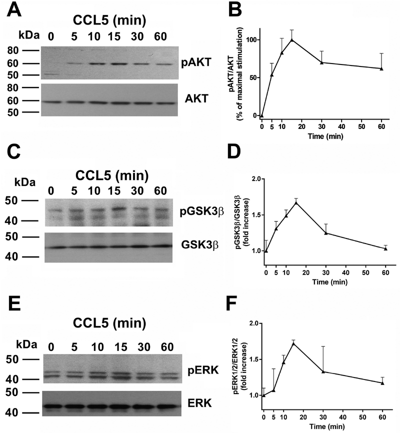
Figure 2. CCL5 increases phosphorylation of signaling proteins.
Differentiated SH-SY5Y cells were exposed to CCL5 (100 nM) for the indicated time points and lysates were prepared. An equal amount of proteins was loaded on a gel, transferred and immunoblotted with an antibody against (A) pAKT, (C) pGSK3β or (E) pERK1/2. Blots were stripped and reprobed with antibodies against AKT, GSK3β or ERK1/2. B, D and F: relative levels of phosphoproteins were calculated by optical density as described in Materials and Methods. Data are the mean + SEM of three independent samples for each time point.

