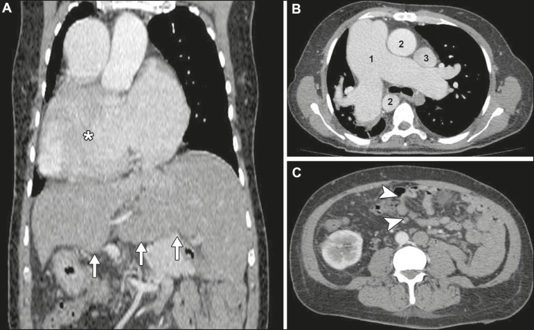
Figure 2.
Contrast-enhanced CT scan. A: Coronal thoracoabdominal CT scan showing dextrocardia (asterisk) and a centrally located liver (arrows). B: Axial chest CT demonstrating enlargement of the right-sided pulmonary trunk, which measured 5.8 cm in its largest diameter (1), right-sided aorta (2) and left-sided superior vena cava (3). C: Axial abdominal CT showing abnormal intestinal rotation (arrowheads)—the entire small bowel is positioned to the left of the midline.

