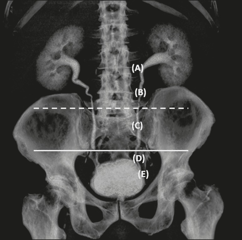
Figure 2.
Three-dimensional reconstruction of an MDCT examination of the urinary tract. Note the ureteral anatomic division: the UPJ (A); the proximal ureter (B); the mid-ureter (C), between the upper border of sacroiliac joint (dashed line) and its lower border (solid line); the distal ureter (D); and the UVJ (E).

