Figure 1.
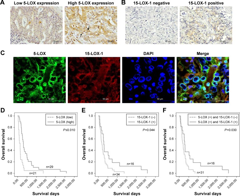
The expression and localization of 5-LOX and 15-LOX-1 in CCA tissues and their correlation with prognosis.
Notes: (A) Representative images of IHC demonstrating the high and low expression of 5-LOX in CCA tissues. Positive inflammatory cell staining was observed as indicated by the black arrow (magnification, ×400). (B) The negative and positive signals of 15-LOX-1 in CCA tissues were determined by IHC staining (magnification, ×400). (C) The co-localization of 5-LOX and 15-LOX-1 immunofluorescence staining in CCA tissues was evaluated (magnification, ×400). (D–F) Survival analysis of 5-LOX, 15-LOX-1, and combination of 5-LOX and 15-LOX-1 expression in each patient with CCA by the Kaplan–Meier method.
Abbreviations: CCA, cholangiocarcinoma; IHC, immunohistochemistry; 5-LOX, 5-lipoxygenase; 15-LOX-1, 15-lipoxygenase.

