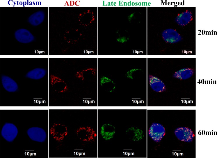
Fig 6. Surface binding and internalization process of ADC by confocal laser scanning microscopy.
The BT474 cells were transduced with BacMam 2.0 CellLight Late Endosomes-RFP and BacMam GFP Transduction Control to stain late endosomes (green) and cytoplasm (blue), respectively. The DM1-ADC4 (red) was labeled with Alexa Fluor 647 and stained cells at 2 μg/mL in PBS buffer containing 10% inactivated goat serum and 1% BSA. The cytoplasm, late endosome, and ADC were excited by lasers with wavelength of 488 nm, 543 nm, and 633 nm.

