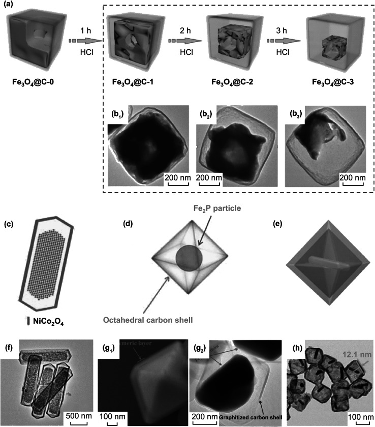
Fig. 3.
a Schematic illustration YS Fe3O4@C nanobox following 1-, 2- and 3-h etching time. The TEM images (b1, b2, b3) of Fe3O4@C nanobox are shown in the dashed box corresponding to 1-, 2- and 3-h etching, respectively. Reprinted with permission from Ref. [84]. Graphical illustrations of c YS nanoprism of Ni–Co mixed oxide, d YS octahedral Fe2P@C, and e YS octahedral Au nanorod@Cu7S4. f TEM image of YS Ni–Co mixed oxide nanoprism. g1 SEM image and g2 TEM octahedral Fe2P@C. h TEM octahedral Cu7S4 shell with Au nanorod yolk. Reprinted with permission from Refs. [80, 82, 88]

